Artificial Intelligence helps us detect coronary heart disease
Every year more than 17.5 million people die worldwide as a result of cardiovascular diseases (2020 data).
For this reason, detection at the earliest and most treatable stage is important. Current screening tests may include laboratory tests to evaluate blood and other fluids, genetic tests that look for inherited genetic markers associated with the disease, and imaging tests that produce pictures of the inside of the body.
Depending on the results of initial screening tests and the presence of risk factors for coronary artery disease, additional tests may be recommended, including:
Electrocardiogram: measures the electrical activity of the heart and reveals information about heart rate and rhythm.
Stress test: involves walking on a treadmill or pedalling a stationary bicycle at increasing levels of difficulty, while heart rate and rhythm, blood pressure and the electrical activity of the heart are monitored.
Cardiac CT for calcium scoring: examines the coronary arteries to measure the amount of calcium in the coronary arteries, an indicator of the amount of plaque in the arteries.
Echocardiogram, which uses ultrasound to create moving pictures of the heart.
Myocardial perfusion scan (also called a nuclear stress test): a small amount of radioactive material is injected into the patient and collects in the heart. A special camera takes images of the heart while the patient is at rest and after exercise to determine the effect of physical and emotional stress on blood flow through the coronary arteries and heart muscle.
CT computed tomography coronary angiography (CTCA): uses computed tomography (CT) and an intravenous contrast medium (dye) to create three-dimensional images of the coronary arteries, and determine the exact location and degree of plaque build-up.
Catheter angiography: takes pictures of the blood flow through the coronary arteries, allowing the physician to see any blockage or narrowing of the coronary arteries (stenosis). During catheter angiography, a thin plastic tube (called a catheter) is inserted into an artery through a small incision in the skin. Once the catheter is guided into the heart, a dye is injected through the tube, and images are captured using X-rays.
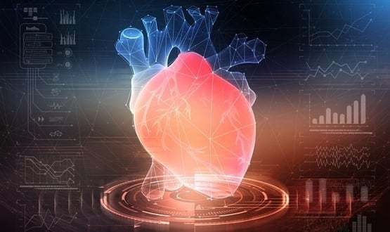
Predicting heart attacks using AI

Source: Digitalhealth
Usually, when someone suffers from chest pain, a CT/CT scan of the coronary arteries is usually done to check for blocked or narrowed areas. In about 70 per cent of cases nothing is found, but some of these people still have coronary heart disease, which could lead to a heart attack.
Doctors do not have the means to detect these cases so that preventive treatment can be applied.
This is where CaRi-Heart® comes in, a technology launched by Caristo Diagnostics, whose founder is Professor Charalambos Antoniades, a senior research fellow at the British Heart Foundation (BHF).
This new technology uses Machine Learning and Deep Learning applied to routine hospital CT scans, finding a specific combination of changes that reveal areas of inflammation and scarring in the fatty tissue surrounding the coronary arteries. Showing that detecting inflammation and scarring significantly increases our ability to predict a future heart attack up to five years before it occurs.
The way they do this is by obtaining, what they call, a Fat Attenuation Index Score® (FAI-Score®), which accurately measures inflammation in the blood vessels in and around the heart. In their study they found that patients with an "abnormal" FAI had a high chance of dying from a heart attack within the next 10 years and that patients who were initially considered low risk had an elevated risk of having a heart attack after having their CT scans reanalysed with this new technology.
It has recently received CE mark accreditation and can now be used by doctors in the UK and Europe.
AI is undoubtedly going to be increasingly present in our lives and the field that will benefit the most is healthcare.
We are at the beginning of the so-called fourth industrial revolution.


